Online supplement"Identification of peripheral neural circuits that regulate heart rate using optogenetic and viral vector strategies", P. Rajendran, R. Challis, C. Fowlkes, P. Hanna, J. Tompkins, M. Jordan, S. Hiyari, B. Gabris-Weber, A. Greenbaum, K. Chan, B. Deverman, H. Munzberg, J. Ardell, G. Salama, V. Gradinaru, K. Shivkumar, bioRxiv preprint (Oct. 2018) |
Online volume data viewer:Click on the images below to interactively view the confocal stack in the browser. Image data has been downsampled ~4x from original resolution. For best viewing, we recommend selecting "No Shadow" and adjusting the contrast and transfer function to best visualize nerve fibers and tissue anatomy.
|
|
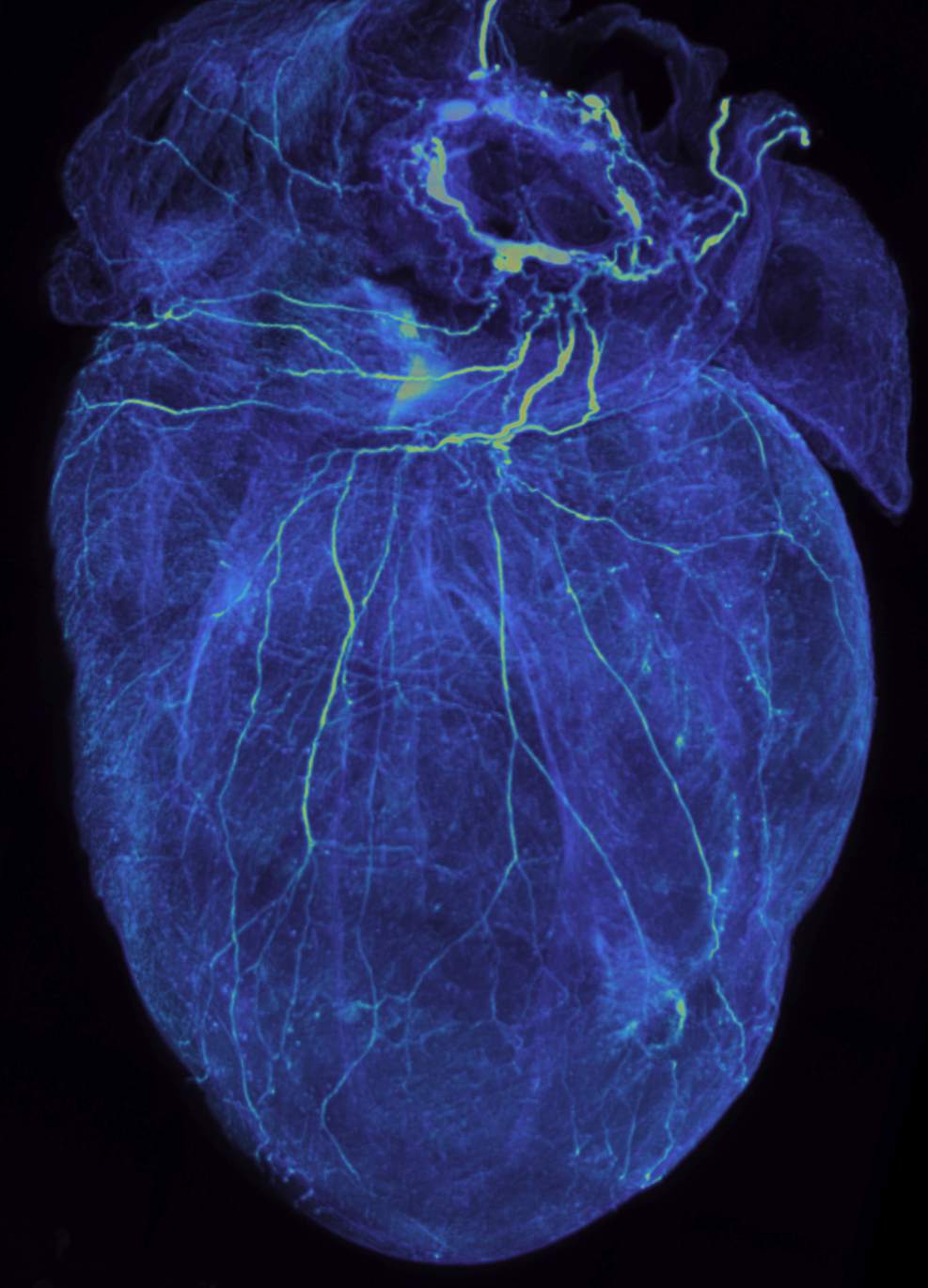
Mouse Heart, dorsal surface [78MB] 3D confocal stack (1200 µm z-stack) of the dorsal side of cleared mouse heart rendered transparent with the iDISCO protocol and stained with PGP9.5 to reveal innervation pattern.
|

|
Mouse Heart, whole [447MB] 3D confocal stack of the whole cleared mouse heart rendered transparent with the iDISCO protocol and stained with PGP9.5 to reveal innervation pattern. Image quality is highest on the ventral surface which was closest to the objective during imaging.
|
Code for widefield tracing, analysis and visualization:Code for analysis and visualization of tracings [scripts] [neuTube] |